banieres_EFER_01
Dropdown Four CSS-Menu
 
Index >  Technique  > Endoscopic assessment
Endoscopic assessment

Careful examination of any endobronchial lesion seen during fiberoptic bronchoscopy is essential. Care must be taken to avoid causing bleeding or edema which can lead to respiratory distress. The exploration of the tracheobronchial tree must be systematic and complete. It is always best to study first the bronchial tree which is pre-supposed free of disease. Any abnormality should be recorded, including anatomic variants which are quite commonly seen, such as a tracheal bronchus, a bi-furcated upper lobe bronchus, or abnormal peripheral distribution. Each bronchus must be properly identified and named.

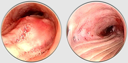
The endoscopist should note the appearance of any lesions, their intrinsic or extrinsic nature, and their location with respect to the carina, vocal cords, etc. The degree of obstruction should be noted in terms of airway patency, and length of involvement as well as the presence or absence of distal secretions. Assessing the apparent vascularity of a tumor is also important because it might help determine the therapeutic approach to a given lesion.


Histologic assessment


Biopsies are almost always performed at the time of fiberoptic bronchoscopy except in high-risk patients with respiratory compromise (high-grade tracheal tumor, bilateral involvement, or recurrence following pneumonectomy).

In these cases, sampling should be postponed until rigid bronchoscopy can be performed.

Multiple, large specimens are necessary to ensure accurate histologic diagnosis of most lesions.

Brush specimens for cytology are not as reliable as biopsy samples.


Histologic analysis of biopsy specimens is mandatory for establishing a diagnosis and prognosis.

Most lesions can be classified as inflammatory or tumoral. Inflammatory lesions often lead to tracheal stenosis and may be associated with respiratory compromise.

 
move up
move down
previous
next
https:/www.high-endrolex.com/30